WELCOME

Op.Dr. Taner CEYLAN Pediatric Urologist


CREATE APPOINTMENT

What I Provide Treatment

Bedwetting Problems
Urgent urge to urinate (urinary urgency)
Adolescent Varicocele (Varicocele in Varicocele)

Treatment of urological pediatric diseases

Endoscopic ureter stone treatment

URS (Incisionless Ureteral Stone Operation)

RIRS (Incisionless Kidney Stone Operation)

Kidney Ultrasound

Bladder stone
(open surgery)

Services

Endoscopic ureter stone treatment


Endoscopic ureter stone treatment is a method used to treat stones in the ureter, one of the tubes that carries urine from the kidneys to the bladder. It is performed by inserting an endoscope into the ureter. Endoscopic ureteral stone treatment is usually used in the following cases:

  • If the size of the stone is 1 cm or less
  • If the stone is located in the lower half of the ureter
  • The stone is lodged in the ureter or the ureteric opening is blocked
Endoscopic ureteral stone treatment is usually performed under local anesthesia and does not require hospitalization. During the procedure, an endoscope is inserted into the ureter and the stone is visualized. Then, using a special instrument, the stone is broken into pieces or removed completely. After the procedure, a stent may be placed in the patient's urinary tract. Endoscopic ureteral stone treatment is usually safe and effective. However, in some cases, complications can occur during the procedure, for example bleeding or infection. Therefore, as with any medical procedure, endoscopic ureteral stone treatment should be carefully evaluated before being recommended by a doctor.

Curiosities in Endoscopic Ureteral Stone Treatment


How is endoscopic ureteral stone treatment performed?

Endoscopic ureteral stone treatment is performed by inserting an endoscope into the ureter. The endoscope is used to visualize the stone inside the ureter. Depending on the size and location of the stone, the stone can be broken into pieces or removed completely.

Is endoscopic ureteral stone treatment painful?

Endoscopic ureteral stone treatment is usually performed under local anesthesia and no pain is felt. However, mild pain or discomfort may be felt after the procedure. This usually passes within a few days.

How long does endoscopic ureteral stone treatment take?

Endoscopic ureteral stone treatment usually takes 30-60 minutes. However, the time may vary depending on the size and location of the stone.

What complications can occur during endoscopic ureteral stone treatment?

Complications can rarely occur during endoscopic ureteral stone treatment. These can include bleeding, infection, narrowing of the ureter or problems with stent placement. However, the risk of these complications is very low if the doctors performing the procedure are experienced and perform it carefully.

How is the recovery period after endoscopic ureteral stone treatment?

After endoscopic ureteral stone treatment, a stent may be placed in the patient's urinary tract. The stent is used to keep the urinary tract open and speed up recovery after the procedure. Patients can usually return to normal activities within a few days, but may be advised to avoid certain activities until the stent is removed. After the procedure, it is important to follow the instructions given by the doctor.

Ureteroscopy - Rigid Ureteroscopy (URS)


The treatment of kidney stones performed with rigid ureteroscopy is referred to as URS. There are various alternative treatments for kidney stone treatment. We have previously discussed PNL, RIRS, and ESWL treatments. The choice of treatment type varies depending on the size and location of the stone, but these three alternatives are used interchangeably. With these methods, the stone inside the kidney is either removed from the body or expected to be expelled with urine. Sometimes a kidney stone may pass out of the kidney spontaneously or as a result of a procedure, but it may get stuck in the urinary tract due to its shape or size. In such cases, the treatment of choice is URS. In URS, no incision is made in the patient's body. The patient's urinary tract is accessed with a device called a ureteroscope. The kidney stone is intervened with a stone-breaking instrument and is broken into pieces. Large pieces are removed from the body during the procedure with the help of equipment called a basket. The remaining stone dust is expected to be expelled through the urinary tract.

How Is URS Performed?

URS is performed in an operating room environment and is often done under general anesthesia. Once the rigid ureteroscope is inserted into the patient's urinary tract, the stone is reached. At this point, the kidney stone is broken into pieces using a laser or pneumatic stone-breaking equipment. Subsequently, large stones are removed from the body using an instrument called a basket. Small stones can easily be passed through the urinary tract. The duration of the procedure varies depending on the size and hardness of the stone, typically taking between 30-60 minutes. At the end of the operation, a thin plastic stent called a double-J (DJ) stent is placed in the ureter, which has one end in the kidney and the other end in the bladder. The reason for placing this stent may vary. If the stone is adhered to the ureter walls, if the procedure has been lengthy, if there is damage to the ureter during the procedure, or simply as a precaution, a DJ stent may be inserted. The stent is usually removed 1-6 weeks after the URS operation, with close monitoring through cystoscopy. It is essential to ensure that it does not remain in the body for more than 2 months.

Who Can Undergo URS?

URS treatment is the first choice when a kidney stone is located in the urinary tract. Kidney stones sometimes move out of the kidney on their own but can become stuck in the urinary tract due to their size. Additionally, after ESWL treatment, if a stone cannot be completely fragmented and exits the kidney, it may become trapped in the urinary tract. In such cases, URS is performed.

Who Is Not Eligible for URS?

If the stone is located inside the kidney's cavities, this method is not applied. It is not performed on patients with active urinary tract infections, severe bleeding problems, or orthopedic issues in their legs. Moreover, it cannot be performed on patients who are not suitable for anesthesia since the procedure is performed under general anesthesia.

Advantages of URS

  • No incision is made on the patient's body. The procedure is carried out through a very thin tube inserted into the urinary tract.
  • Patients can be discharged on the same day.
  • Within 2-3 days, patients can usually return to their normal lives.

What to Expect After URS?

After the procedure, patients may experience:

  • Burning sensation during urination
  • A small amount of blood in the urine
  • Frequent and urgent need to urinate. These symptoms often resolve on their own a few days after the procedure.

When Should I Call My Doctor After the Procedure?

  • Fever exceeding 38.5°C that doesn't subside
  • Burning sensation during urination is normal. However, if it remains severe or does not subside even after a few weeks,
  • If you are unable to urinate
  • If there is a significant amount of blood in the urine
  • If you continue to experience side pain, you should contact your doctor.

What Is the Difference Between RIRS and URS?

RIRS and URS are quite similar procedures. In both methods, no incision is made in the patient's body. Access is made through the urinary opening. In URS, a rigid ureteroscope is used, while in RIRS, a flexible ureteroscope is used. RIRS can be described as an advanced version of URS. URS is used to treat stones located in the urinary tract, while RIRS can treat stones inside the kidney.

RIRS (Non-incision Kidney Stone Surgery)


What Is RIRS?

RIRS is the newest method used in the treatment of kidney stones. Kidney stone disease is quite common in our society. Individuals often visit urology clinics suspecting conditions such as flank pain and burning during urination. Physical examination and ultrasound results determine whether there are stones in the kidney and also monitor the size and location of the stone. Although there are many different methods for kidney stone treatment, before RIRS, for large stones (over 2 cm), the only option was PNL closed kidney stone surgery and open surgery. As known, PNL involves making a small incision in the patient's body to reach the kidneys and intervene in the stone. However, with RIRS, it became possible to intervene in kidney stones up to 3.5 cm without making an incision in the patient's body.

What Is RIRS Kidney Stone Treatment?

RIRS is performed in the operating room under general anesthesia. A highly flexible ureteroscope is inserted through the urinary tract to reach the stone in the kidney. The stone is broken into small pieces and removed from the body. Unlike PNL, the most important advantage of this method is that the procedure can be performed without making an incision in the body.

For Which Stones Is RIRS Applied?

  • Small, medium, and large stones up to 3.5 cm
  • Overweight patients
  • Patients with multiple stones
  • Patients with stones in both the kidney and ureter
  • Stones for which ESWL treatment did not yield results
  • Patients with stones in both kidneys

Advantages Compared to Other Kidney Stone Treatments

  • One of the most significant advantages compared to PNL is that there is no need to create an incision in the body. This is a crucial advantage that shortens the hospitalization time and reduces the risk of infection and bleeding.
  • It is possible to be discharged 24 hours after the procedure. This period can extend up to 3 days in PNL.
  • Compared to ESWL, the most important advantage is the ability to apply it to larger stones. ESWL can be applied to stones smaller than 2 cm, whereas RIRS can be used for stones up to 3.5 cm.
  • In ESWL, it is expected that all the broken stones will be excreted through the urinary tract. This may pose a risk of blockage in the urinary tract depending on the size of the intervened stone. However, during the RIRS procedure, large stones are removed from the body. Small debris should be excreted with urine.
  • It can be applied to patients using blood-thinning medications.
  • For patients with congenital kidney anomalies, this method is the primary choice.

Disadvantages Compared to Other Methods

  • RIRS was initially tried on small stones and successful results were achieved, but as technology developed, it was applied to larger stones up to 3.5 cm. Since breaking large stones takes more time, the operation may be prolonged. Consequently, the ureter may be damaged, and stricture may occur.
  • Due to its relatively recent status, it can be performed by a limited number of centers and surgeons.

After RIRS

A double J stent may be inserted during the procedure. One end of the stent is attached to the kidney, and the other end is connected to the bladder to prevent ureteral obstruction and facilitate urine flow. The stent must be removed within 1.5-3 months. A catheter may be placed for overnight monitoring. The catheter is removed before discharge. Patients are generally discharged the day after the procedure.

RIRS in Kidney Stone Treatment

The use of RIRS in kidney stone treatment was first attempted in the early 2000s, mostly for small stones. With advancing technology, it has been applied to larger stones and more patients, resulting in successful outcomes. Today, RIRS can be used to intervene in stones up to 3.5 cm in size. The number of surgeons who have received training and gained experience in this regard is limited. However, considering the advantages of RIRS over other kidney stone treatments, it is expected that the method will be used by many more centers in the coming years.

Kidney Ultrasound


What Is Kidney Ultrasound?

Ultrasound is an extremely practical imaging method today, thanks to advanced technology. Renal ultrasound, also known as kidney ultrasound, facilitates the visualization of issues in patients' kidneys and their surroundings. The purpose of this ultrasound is to assist in the visualization of problems occurring in the kidneys and the surrounding tissues. In kidney ultrasound imaging, not only the kidneys but also the bladder and the channels between the kidney and the bladder can be easily examined. It is a widely used imaging system in today's medical practice, essential for accurate early diagnosis and treatment. Ultrasound imaging allows the diagnosis of health problems in the abdominal area, including the kidneys. It enables the diagnosis of issues such as kidney stones, masses, and other conditions in the kidneys, making early treatment possible. Regular ultrasound imaging is recommended for patients who may have a genetic predisposition to kidney problems, as early diagnosis is crucial.

Why Is Kidney Ultrasound Performed?

Early diagnosis is of great importance in kidney ultrasound imaging. The purpose of this imaging system, which aims for early diagnosis and treatment, is to detect urinary tract infections and observe the presence of masses, stones, or sediments in the kidneys. Kidney issues can become severe health problems if not detected early, and regular kidney ultrasound imaging is crucial for early diagnosis. Untreated and undiagnosed kidney disorders can lead to kidney failure if they progress. Therefore, kidney ultrasound imaging plays a significant role in preventing such complications. The ultrasound devices used by expert physicians for disease diagnosis are continually advancing, providing the necessary tools for early diagnosis. With the advancement of technology, these imaging procedures have become easy to perform, and patients usually do not experience any pain or discomfort during the procedure. It is important for the patient to have a full bladder during the imaging, which is achieved by drinking plenty of water to ensure clear and accurate results.

Open Bladder Stone Surgery


Bladder stones are removed through open surgery and other treatment alternatives. Bladder stones can form due to nerve damage, bladder outlet obstruction, prostate enlargement, or urinary tract infections, and they can also occur for congenital reasons. When their symptoms are observed, it is crucial to seek treatment, as they can lead to various obstructions and kidney damage if left untreated.

When Is Open Bladder Stone Surgery Performed?

Open bladder stone surgery is usually preferred when the closed surgical method cannot be applied. If the bladder stone is very large, if it can only be treated with an open method due to an underlying prostate enlargement, or if a diverticulum surgery needs to be performed together, open surgery is used. Open bladder stone surgery is used to treat bladder stones, and it also checks for any urinary tract obstructions. Obstructions can be eliminated using open or closed techniques depending on the case. Open surgery is performed less frequently today, but it is still chosen when necessary.

How Long Does Bladder Stone Surgery Take?

Open bladder stone surgery is typically completed in about 1-2 hours. The duration may vary depending on the size of the bladder stone, additional procedures, and the patient's overall health. Thanks to advanced surgical techniques, success rates have increased. Proper preoperative planning helps eliminate existing surgical risks. As with any surgical intervention, there can be side effects after bladder stone surgery. Pain and discomfort in the area are among the most common issues. Following a successful bladder stone surgery, the patient will quickly recover from their complaints and resume their daily life. If you would like to learn more about open bladder stone surgery and our practices, please contact our clinic.

Treatment of Pediatric Urological Diseases


What is Pediatric Surgery?

In our clinic, we provide polyclinic follow-up and treatment for all surgical problems related to the digestive system, urinary tract, thoracic cavity excluding the heart, and head and neck anomalies in children from the prenatal period to the newborn period up to the age of 16. Surgical procedures are performed using closed (minimally invasive, laparoscopic, thoracoscopic, endoscopic) or open methods, depending on the type of the disease. With the knowledge, skills, and expertise of our team, as well as advanced technological resources, we use advanced technology both in the diagnosis and treatment areas.

What is Pediatric Urology?

In Pediatric Urology, urinary problems in children, bedwetting, or urinary incontinence issues are diagnosed and treated in our high-tech pediatric urology diagnosis and treatment center. In addition, our clinic, which provides services with a 24/7 expert staff, is aware of the importance of a multidisciplinary approach in pediatric patients and pediatric surgery. We work together in terms of preoperative preparation, painless surgical procedures, and follow-up with pediatric anesthesia. With the multidisciplinary council and programs established within our hospital, we collaborate with all branches, especially with pediatric health and diseases clinic physicians, to achieve national and international success in the diagnosis, diagnosis, and treatment of our patients. The presence of fully equipped neonatal and pediatric intensive care units and all sub-branches of pediatric health and diseases is one of the most important components of perfection and high success in postoperative care of complex surgical patients.

Multidisciplinary Programs


Here are some of the multidisciplinary programs we offer:

  • Gender Development Disorder Program
  • Pediatric and Adolescent Thyroid Cancer Program
  • Tube Feeding Program in Children
  • Chronic Abdominal Pain Program in Children
  • Pediatric Inflammatory Bowel Disease Program
  • Pediatric Hepatobiliary Diseases Program
  • Constipation Program in Children
  • Gastroesophageal Reflux Program in Children
  • Pediatric Solid Abdominal Tumors Program
  • Hemangioma and Vascular Malformation Program in Children
  • Spina Bifida Program
  • Nocturnal Enuresis and Pediatric Urinary Problems Program
  • Hydronephrosis Program in Children
  • Torticollis Program
  • High-Risk Baby Program
  • Rare and Difficult-to-Diagnose Diseases Program

Photo

Op.Dr. Taner CEYLAN


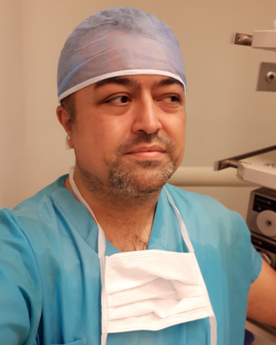
Dr. Taner CEYLAN graduated from Ankara Private Yükseliş College in 1991 and Hacettepe University Medical School in 1997. Faculty of Medicine. In 1998, he started to work as an assistant at Ankara Numune Training and Research Hospital and In 2002, he became a Urology specialist. As a urologist, he worked at Ankara Numune Hospital and Ankara Yenimahalle He worked at the State Hospital.

FEAPU

In 2015, he successfully passed the Sub-Subspecialty Examination in Medicine and graduated from Hacettepe He started his subspecialty residency in Pediatric Urology at the University Department of Urology. In 2019, he started his residency in Pediatric He became a subspecialist in urology. In the same year, he passed the European Pediatric Urology exam in Leuven-Belgium. Certificate of Expertise (FEAPU).

As a Pediatric Urology specialist, he started his compulsory service at Hatay Antakya State Hospital. and completed his compulsory service obligation in Ankara Dr. Sami Ulus Children's Hospital in 2022. Dr. Taner CEYLAN has been continuing his professional life only in the field of pediatric urology since 2016 and diagnosis of congenital and acquired urologic pathologies in childhood and medical and surgical is interested in treatment. Dr. Taner CEYLAN, who speaks fluent English and intermediate German, is married and has 2 sons.

CONTACT

Contact us for any questions or appointments!